
IBIF Resource List
IBIF hosts five primary imaging instruments, as well as data analysis workstations as outlined below.
This Leica TCS SP8 microscope is well-suited for small animal imaging and electrophysiology experiments.
- Lasers: 405, 488, 514, 552, 633, and a Mai Tai 680-1040 nm tunable Ti:S
- Super HyD detectors with Becker and Hickl Time Corrleated single photon counting system
- Capable of Multiphoton, SHG, spectral imaging, and standard confocal imaging

This spinning-disk confocal can image at speeds up to 1,000 frames per second, making it our only confocal microscope that can image in “real time”.
- Equipped with Yokogawa spinning disk and back-illuminated Photometrics’ Kinetix sCMOS camera for ultrasensitive detection
- Lasers: 405, 488, 561, and 640 nm
- Super-resolution capable with Gataca’s Live SR
- Tokai Hit stage-top incubation system
- More than 10 objective options available
- Ideal for live cell imaging in real time as well as for longer-term time lapse imaging

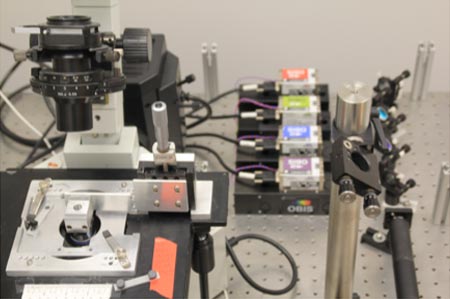
This through-the-prism TIRF system was built in-house on an Olympus’ inverted microscope body.
- Lasers: 405, 488, 552, and 647 nm
- Image capture with Andor iXon EMCCD camera
- Ideal for imaging of samples on slides that require high speed and/or high sensitivity
This widefield imaging platform with built-in incubation enables multi-day imaging experiments.
- Imaging options: fluorescence, brightfield, phase contrast
- Stage inserts allow users to image with microscope slides or well-plates
- Users have successfully completed 3 – 5 day imaging experiments that were fully-automated

This widefield microscope with sCMOS camera is ideal for basic imaging and sample inspection. Image capture with Hamamatsu Orca Flash Capable of FURA-based imaging experiments.
Software Supported
IBIF encourages users to work with vendor-supported software or the FIJI version of ImageJ. In addition, IBIF has a dedicated workstation for the analysis version of Nikon’s NIS Elements, which includes the powerful GA3 analysis platform. If a user has questions regarding analysis software, please ask a member of the team.
- ImageJ (Fiji version)
- Source/Vendor: NIH
- Purpose: Data Analysis
- LAS X
- Source/Vendor: Leica
- Purpose: Data Acquisition and Analysis
- MetaXpress
- Source/Vendor: Molecular Devices
- Purpose: Molecular Devices
- NIS Elements
- Source/Vendor: Nikon
- Purpose: Data Acquisition and Analysis
- MATLAB
- Source/Vendor: Mathworks
- Purpose: Data Analysis
- Python
- Source/Vendor: N/A
- Purpose: Data Analysis
- R/RStudio/Posit
- Source/Vendor: N/A
- Purpose: Data Analysis
Some of these software packages are readily available for individuals to download or access online. All of these packages are available on one or more workstations within the core facility, but users are billed for using the core’s analysis workstations at an hourly rate.
Additional Resources
Additional laser scanning confocal microscopes and image analysis tools are available through the Dr. Richard J. Bellucci Translational Hearing Center. More information about our sister facility can be found under the “Advanced Imaging” section on this webpage.
